More permeable than expected: Researchers have found out more about newly discovered mini-canals in our skull bones. According to them, these connections between the brain and bone marrow contain immune cells that are unique in their composition and response to disease. It was also shown that inflammatory processes in the brain can also be detected on the surface of the skull. This opens up new avenues for the early detection and treatment of neurological diseases, according to the team in the specialist magazine Cell.
Our skull bones have long been considered a kind of helmet that protects the brain from external influences, but has no direct connection to the inside itself. But recently, researchers discovered tiny channels in the skull bone that allow immune cells to travel directly from the skull’s bone marrow to the brain. In addition, it turned out that a previously unknown, fourth meninges acts both as a protective layer and as a base for immune cells.
Search for traces in the skull bone
Building on these findings, a team led by Zeynep Ilgin Kolabas from the Helmholtz Zentrum München has now gained further insights into the structure of the cranial tubules and the composition of the immune cells found in them using new methods. The researchers placed a special focus on inflammatory processes that occur, for example, after a stroke and lead to the activation of numerous immune cells.
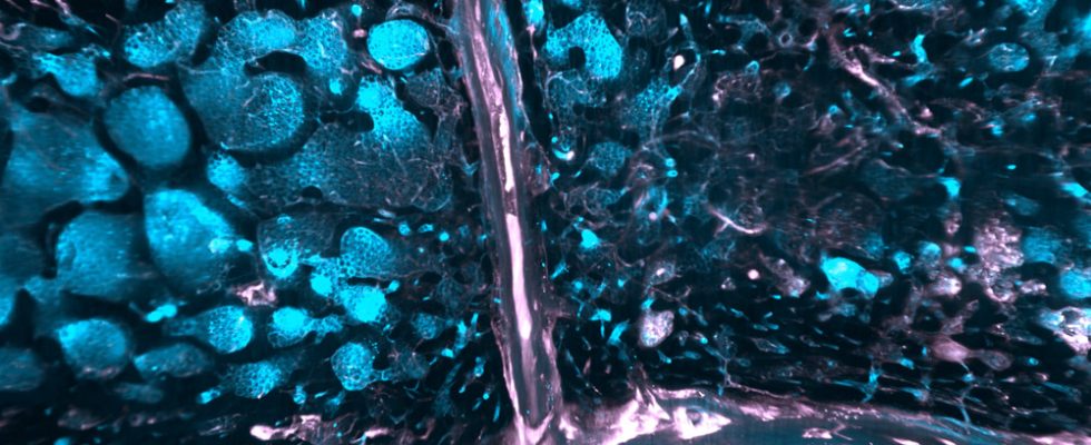
In a first step, Kolabas and her team induced a stroke in mice, then killed the animals and made their tissue transparent with a special solution. This “tissue clearing” enabled them to analyze the skulls of the mice with high-resolution 3D imaging and gain insights into where which cells accumulate as a result of the stroke.
The team also analyzed skull bones from human donors in a similar way, making the channels between the bone marrow and the brain visible. In addition, the researchers examined the composition of the RNA transcripts and the proteins in the skull and other bones in both mice and human bones.
Unique defense cells
The analyzes revealed: Unique neutrophilic immune cells are released in the marrow and tubules of the cranial bone in response to inflammation of the brain. This type of white blood cell is not found in the marrow of other bones in the body, comparative analysis has shown. It was also shown that the skull bone differs significantly from all other bones in the body in terms of its gene activity and proteins.
The investigations on human skull samples also provided interesting details on the cranial tubules: “The lumen of these canals are lined with a layer of fibroblastic cells, which are also known as antigen-presenting cells,” the researchers report. These immune cells could serve as sensors, sampling the invading cerebrospinal fluid for possible signs of infection or inflammation.
Fat-filled tubes and disease-indicating proteins
The tissue clearing also revealed that, unlike mice, the cranial canals in humans are filled with fats and other lipids. According to Kolabas and her colleagues, this lipid filling could facilitate the transport of immune cells through the cranial tubules. At the same time, the fatty substance could also serve as an energy source for bone marrow stem cells.
Using brain scans using positron emission tomography (PET scans), the researchers also found that the inflammatory processes in diseases such as Alzheimer’s or strokes are associated with specific brain signals. “In the brain, the TSPO protein is noticeably upregulated in such neuroinflammatory processes,” they report. This can be demonstrated using PET. To their surprise, they found the signal from these proteins not only in the brain itself, but also in cells of the overlying cranial bone.
Recognize brain inflammation in the skull
“Our results suggest that the connection between the skull and the brain is far more complex than previously assumed,” says Kolabas. At the same time, the newly discovered reaction of the skull to processes inside the brain also opens up new possibilities for the diagnosis and treatment of brain diseases. In the future, encephalitis could possibly be monitored simply by scanning the surface of the affected person’s head.
“This could lead to more effective monitoring of diseases such as Alzheimer’s and stroke and possibly even help to prevent the onset of these diseases through early detection,” says Kolaba’s colleague Ali Ertürk. (Cell, 2023, doi: 10.1016/j.cell.2023.07.009)
Source: Helmholtz Zentrum München German Research Center for Environmental Health (GmbH)