Researchers are developing a new approach to proteomics that enables the long-awaited single-cell resolution on intact tissue
The function and protein composition of individual cells depend on what resources are available in their immediate surroundings. Thanks to a new approach by the research team led by Matthias Mann, director at the Max Planck Institute for Biochemistry in Martinsried, it is now possible for the first time to analyze this protein composition with a view to the environment of individual cells. By combining two new methods from the research department, they were able to functionally record the proteome of individual cells with high spatial resolution.
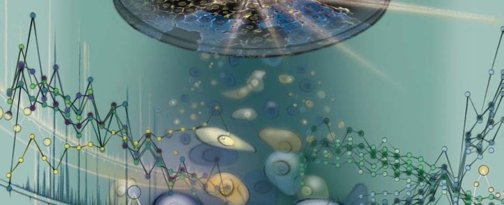
© Juliet Percival Illustration of proteome analysis of a single cell in a tissue context. © Juliet Percival
Tissues are made up of many different cell types, each of which performs specific functions in our body. Even within such a cell type category, however, the function of each individual cell depends on the environment it is in, for example whether there are blood vessels near it or what other cells are around it. Are there many usable nutrients? How much oxygen is available to the cell? Which hormonal signals affect them?
This connection between function and location is particularly pronounced in the liver. The liver, as the body’s central metabolic organ, consists of 85 percent of one type of cell called hepatocytes. These cells perform various metabolic tasks in our body, such as storing glucose or detoxifying our blood. The respective proteins within the cells are of crucial importance in these metabolic processes. However, it has not yet been possible to measure and display the proteome of individual liver cells with a view to the cells’ immediate surroundings.
Further development of deep visual proteomics
Researchers have now closed this gap with a technological milestone. Matthias Mann’s team developed the Deep Visual Proteomics method in 2022 to determine the proteome of different cell types. During analysis with Deep Visual Proteomics, after a tissue sample is taken, cells of a type are identified, extracted and sorted into specific groups. They are then analyzed for their protein composition using mass spectrometry.
“In our new study we have brought two worlds together. We have combined so-called single-cell proteomics with the spatial resolution of Deep Visual Protemics, creating a new approach for single-cell proteomics with spatial resolution. The new approach, Single-Cell Deep Visual Proteomics, now allows us for the first time to measure more than two thousand different proteins of individual cells in intact tissue and thus with spatial resolution,” explains Florian Rosenberger, lead author of the current study. “We are now able to precisely localize hundreds of metabolic pathways,” Rosenberger continued. This can now be used to create protein maps that help to better understand the mechanisms of health and disease.
By combining the two existing methods, the authors were also able to show that neighboring cells sometimes have enormous functional differences. So far, such differences have only been shown for a few proteins. This new approach paves the way for a better understanding of complex tissues such as tumors. This means that individual cells and the mechanisms that lead to resistance in cancer therapies, for example, can now be identified.
“We are reaching historic new territory with the first single-cell proteomics analysis that can be performed directly in the tissue context and the recognition that the same cell types actually perform very different activities depending on where they are located. We are convinced that we already “In just a few years, we can measure almost the entire proteome of a cell, with more than 10,000 proteins in just a few minutes,” says Matthias Mann.
Terms
Proteome: The proteome includes the entirety of all proteins in a living being, a tissue or a cell at a certain point in time. The proteome is highly dynamic and responds to the requirements of the cell, as well as to diseases or environmental influences.
Proteomics: Proteomics is the study of the proteome.
Mass spectrometry: Mass spectrometry is a method for determining the mass and abundance of charged particles.
Deep Visual Proteomics: Deep Visual Proteomics, or DVP for short, is a method that was developed in 2022 in Matthias Mann’s research department. The method combines modern microscopy, machine learning, laser microdissection and ultra-sensitive mass spectrometry with subsequent bioinformatics analyses.