
In the future, the Berlin researchers will have to decide for themselves whether it is worth the trip. An expensive electron microscope has recently been installed on the Buch research campus in the northern tip of Berlin – just before the border with Brandenburg. It will be officially inaugurated on Friday.
“Something like this happens when everyone pulls together,” says Oliver Daumke, structural biologist at the Max Delbrück Center. The MDC, the Charité, the Free and Humboldt Universities as well as the neighboring Leibniz Institute for Molecular Pharmacology have joined forces for this project.
The price for the device alone is five million euros, and with the technology around it it is even eight million euros. The money for this comes from the German Research Foundation. Another 2.9 million was due for the specially constructed cube-shaped building. In addition, there are the costs for operations and the newly built staff. So there was a lot of investment here.
Conditions like in space
The microscope itself has the appearance of space technology: It consists of a wild tangle of cables, tubes and precisely crafted, brightly polished metal that is stuffed into a four-meter-high cabinet.
Inside the machine there are icy temperatures and a high vacuum “like in the stratosphere,” as Christoph Diebold, manager of the company, explains. In this atmosphere, electrons guided by magnets collide with shock-frozen cells. Behind them, a screen detects how much they were deflected from their flight path.
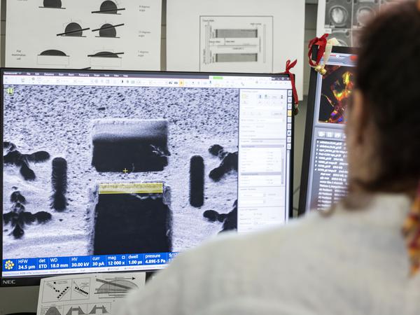
But this is no ordinary cryo-electron microscope, the Charité scientist continues. It is so sensitive that it can image the inner workings of the cell “in the picometer range”. A water molecule is about one hundred picometers in size.
It also has a tilt control: it angles the sample in the electron beam, which illuminates it from different directions. In this way, spatial models of molecules are created from hundreds of thousands of individual images.
A rapid development
The technology for this type of structural biology has developed rapidly over the last 20 years. During this time, the devices learned to resolve a thousand times more details, explains Christian Spahn, researcher at the Charité.
There are around 40 cryo-electron microscopes of various types throughout Germany, spread across a few centers. The technology of this “cryo-EM” closes a gap in biological imaging, says the scientist.
On the one hand, cellular structures would be visible in the light microscope. On the other hand, the spatial structure of isolated proteins is known from crystal studies. “But the nano level in between has been very mysterious for many years,” he says.
Spahn has already used the technique to examine ribosomes from developing brain tissue, but outside the cells. These molecular machines produce all of the body’s proteins. The new device should now make it possible to analyze the particles directly in cells that come from the brains of mice.
There is another device with similar capabilities at the Free University in the south of Berlin. You can only look at the molecules directly in the cell in Buch, says Diebold: “There is no comparable facility in Austria or Germany.”
The electron microscope is a native of Berlin, as it was invented here in 1931 by the future Nobel Prize winner Ernst Ruska together with Max Knoll and further developed at Siemens. After the war, Ruska conducted research at the Fritz Haber Institute in Dahlem.
The patron saint of the “Isolde Dietrich House,” where the new microscope is located, worked with him: the physicist constructed a lens with helium-cooled coils, which is characterized by a particularly stable magnetic field. However, the complex technology did not become established. Conventional magnets are used in Buch, they are cheaper.