Peel our brain with unprecedented precision. This is what Iseult promises, a magnetic resonance imaging (MRI) device designed by the Atomic Energy and Alternative Energies Commission (CEA), whose first anatomical images of the human brain were unveiled on Tuesday April 2 on the Saclay site. In 2021, the 11.7 Tesla of its magnetic field had already revealed the insides of a pumpkin, to mark its entry into an operational phase after twenty years of development. Since then, CEA teams have cautiously moved on to observing their first human subjects.
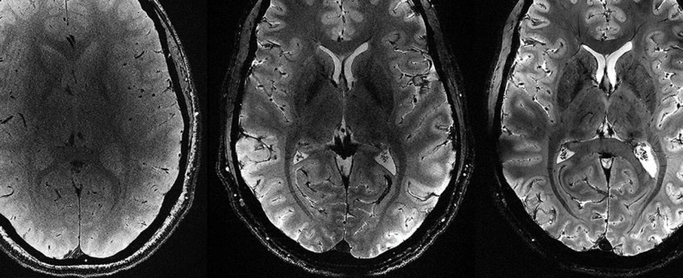
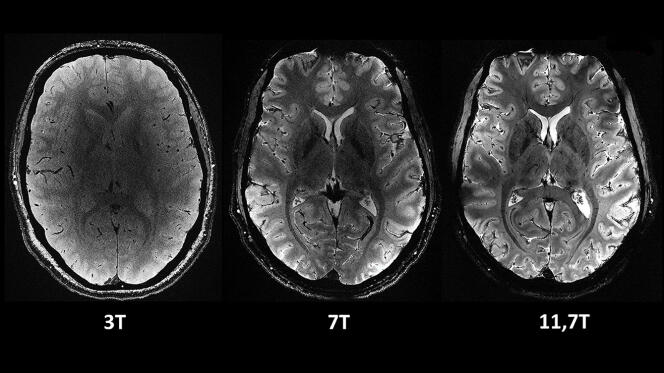
“The images are incomparably clear! », rejoices Nicolas Boulant, responsible for the Iseult project at the CEA. To obtain anatomical images offering the resolution of those provided by Iseult in four minutes of observation – 0.2 mm in the plane and 1 mm in depth – it would be necessary to stay two hours in a hospital quality 3 T scanner. A completely theoretical comparison, since the patient’s movements would then blur the image.
If the transition from pumpkin to man took so long, it is because with Iseult we were entering an unknown territory of cerebral exploration. “It was necessary to prove to the health authorities that such an intensity of magnetic field has no effect on health”, explains Nicolas Boulant. The previous record was 10.5 Tesla on an American machine in Minneapolis. Tests were therefore carried out on 20 healthy adult volunteers to verify balance, cognition, brain tissue temperature, genotoxicity, etc. A nocébo study was even carried out, in which volunteers were subjected, without their knowledge, to a session at zero Tesla, to see if the machine, “which can be intimidating”notes Nicolas Boulant, could induce a psychological bias. “We saw absolutely nothingassures the physicist. It was a crucial step to see if we would be able to really do it an explorer of the human brain. »
“Deciphering the neural code”
A new phase will now open, to continue to refine the acquisition of data according to the different imaging modalities offered by MRI: anatomical data, but also functional data – that is to say visualizing the activated brain areas by this or that cognitive activity. It will also be possible to carry out so-called “diffusion” imaging, which highlights the neuronal bundles connecting the different areas of the brain. The intensity of the magnetic field should also make it possible to detect invisible compounds at lower fields, such as lithium, used in bipolar disorders, and glucose and glutamate, small molecules involved in brain metabolism.
You have 44.61% of this article left to read. The rest is reserved for subscribers.