MRI examinations continue to be of great importance for MS treatment – a comment by Marc Pawlitzki and Sven G. Meuth
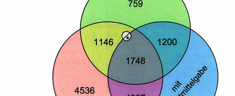
MRI diagnostics continue to have a high diagnostic, but also therapeutic value for the care of MS patients. The diagnostic criteria alone have recently further underpinned the importance of MRI. Already after the first clinical episode, the diagnosis of MS can be made with an appropriate distribution of MS-typical demyelination foci in the initial cranial and spinal MRI. However, there must also be evidence of a contrast medium-enhancing lesion as a criterion for the temporal dynamics. Although this criterion can be replaced by a suitable cerebrospinal fluid finding (detection of so-called oligoclonal bands), this should not lead to the administration of contrast medium being dispensed with. The current data from the MS register of the DMSG shows a pleasing development overall. Because the prompt implementation of the current international recommendations leads to an optimization of previous MRI examinations and thus to a reduction in contrast agent administration that is not necessary. In detail, the administration of contrast media is no longer considered sensible, especially for regular MRI checks, but the current recommendations continue to underline the prerequisites for optimal monitoring using MRI:
1. Standardized MRI protocols, which thus enable good comparability with previous images and
2. Regular cranial MRI scans, particularly to detect insidious disease activity.
Caution is required if one wants to evaluate the success of an initiated immunotherapy by means of an MRI follow-up. Here, three to six months after the initiation of therapy, an MRI examination should be considered as the initial finding (so-called re-baselining), which is then decisive as a comparison for the further MRI controls. The fact that the administration of contrast medium is usually dispensed with as the disease progresses corresponds to previous experience from studies that show an age-dependent decrease in contrast medium-enhancing lesions in MS² . Nevertheless, administration of a contrast agent can be considered, especially if secondary chronic progression is suspected, to identify (still) present inflammatory activity.