Not all Alzheimer’s are the same: Researchers have identified five different Alzheimer’s subtypes – dementia variants with different courses, molecular causes and possible therapeutic approaches. These subtypes can be distinguished by the proteins in the spinal fluid and could help develop more targeted therapies in the future, as the team reports in “Nature Aging”. They also provide an explanation for why current Alzheimer’s medications are only partially effective.
Although Alzheimer’s is the most common neurological disease in old age, neither its causes nor its neurophysiological basis are fully understood. This also hinders the development of effective therapies. The new antibody active ingredients Lecanemab and Donanemab slow down the formation of amyloid plaques in the brains of those affected, but can only slow down the progression of dementia, not stop it. One reason for this could be that Alzheimer’s has inconsistent causes, so that therapies only work on some of these subtypes.
There were already initial indications of such subtypes: the breakdown of brain cells begins in different brain regions depending on the activity of the Alzheimer’s risk genes. Four subtypes were also discovered in the accumulation and spread of misfolded tau proteins.
Spinal cord puncture reveals subtypes
Now there is new, more concrete data on the Alzheimer’s subtypes and their characteristics. It was discovered by a team led by Betty Tijms from the Alzheimer Center Amsterdam when they analyzed the proteins in the spinal fluid of 419 Alzheimer’s patients and 197 healthy controls. Using mass spectrometry and an AI-supported analysis, the researchers examined which of the 3,800 proteins in the CSF were increased or decreased in Alzheimer’s patients and what differences there were.
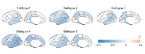
It turned out that 1,058 proteins in the spinal fluid are significantly changed in Alzheimer’s patients – and form striking groupings. “The proteome profiles of Alzheimer’s patients form five subtypes,” report Tijms and her colleagues. These subtypes differ in the type and amount of altered proteins as well as other characteristics. Additional genetic analyzes showed that all five subtypes had a specific pattern of risk gene variants in addition to the well-known Alzheimer’s risk gene APOE4-e4.
Symptoms and progression vary
The five Alzheimer subtypes identified in the CSF are also linked to specific symptoms, dementia progression rates and survival times of those affected. “The subtypes differ, for example, in how they progress from mild cognitive impairment to dementia: subtypes 2 and 5 have the highest risk, subtype 4 the lowest,” the team reports. “Brain scans using magnetic resonance imaging also showed that the subtypes also differ in the extent and location of neuronal degradation.”
There are also differences in the rate of neuronal degradation: patients with subtype 3 had the shortest survival time, on average only around 5.6 years. They also showed a much steeper decline in memory and language tests and the highest levels of misfolded tau proteins, Tijms and her colleagues found. Those affected with subtype 1, on the other hand, survived the longest, around 8.9 years after diagnosis, despite relatively high tau protein levels in the CSF.
Five different causes?
But the new data also provides valuable information about the molecular and genetic causes of the five Alzheimer’s subtypes. As the team determined, subtype 1, for example, is linked to high neuronal plasticity: in the CSF of these patients there are a striking number of proteins that indicate the remodeling and increased activity of brain cells. “Such hyperactive neurons secrete more tau and amyloid and were primarily observed in the area of plaques,” report Tijms and her colleagues.
Subtype 2, on the other hand, is characterized more by immune-specific messenger substances and proteins. Some gene variants in this subtype are also linked to immune processes. “This suggests that an overactive innate immune system is making the disease worse,” say the researchers. In Alzheimer’s subtype 3, they found evidence of RNA dysregulation. A similar disorder has also been observed in frontotemporal dementia.
In subtype 4, patients show a noticeable accumulation of proteins that come from the so-called choroid plexus. This branched network of veins sits on the walls of the cerebral ventricles, produces cerebrospinal fluid and regulates the transport of substances through the blood-brain barrier. In Alzheimer’s patients with subtype 4, this regulation appears to be disturbed, as Tijms and her team report. The breakdown of brain matter is also relatively pronounced in this type. In subtype 5 Alzheimer’s, however, blood-typical proteins in the cerebrospinal fluid indicate that the blood-brain barrier is permeable and can no longer fulfill its protective function.
New starting points for diagnosis and therapy
According to the researchers, these findings open up valuable approaches for better diagnosis and treatment of Alzheimer’s. The differences between the five subtypes could explain why certain therapies only work in some patients. “Given the specific patterns of molecular processes and genetic risk profiles, it is likely that Alzheimer’s subtypes also require specific treatments,” the team writes.
At the same time, the Alzheimer’s subtypes could also explain why, for example, the antibody preparations only cause severe cerebral edema in some patients: those affected with subtype 5 may be particularly susceptible because the blood-brain barrier does not function properly. Knowledge of the subtype could therefore also be important for clinical studies of new active ingredients and therapies. (Nature Aging, 2024; doi: 10.1038/s43587-023-00550-7)
Source: Amsterdam University Medical Center
January 11, 2024 – Nadja Podbregar