Status: 10/26/2022 8:15 p.m
With light sheet microscopy, living cells can be observed – without damaging them. Cancer therapies, for example, can be advanced in this way. Researchers from Jena have now received the Future Prize for the technology.
For a long time, researchers have been trying to stain microscopically small structures in cells with fluorescent dyes. If the cells are already dead, that’s not a problem. But if they are to live on, it is not so easy. Because in order to generate fluorescence radiation, you need laser light, and that kills cells.
At the beginning of the 2000s, scientists developed what is known as light sheet microscopy as a way out: the laser light is not beamed into the sample frontally, but perpendicular to the direction viewed by the microscope. And it is focused so strongly that it only illuminates one layer of the sample, the so-called light sheet. This layer was initially a few micrometers thin, roughly the diameter of a cell. The rest of the sample is not irradiated and is therefore protected.
Only one strip is illuminated
“That is the main advantage of the technology: that the sample survives the imaging and thus comes out of the microscope alive in the end and can continue to live,” said one of the pioneers of the method, Jan Huisken, on Deutschlandfunk in 2019. At that time, he used the method to observe, for example, the development of the heart in the growing zebrafish. Using the light sheet microscope, he was able to see directly when the first heartbeat occurred – on a living object.
But research wanted to go further, reports Thomas Kalkbrenner from Carl Zeiss Microscopy in Jena. “Until now, this has only been used for larger samples – model organisms such as zebrafish or fruit flies. We wanted to develop this for cell biology. And that means the light sheets have to be very thin so that the resolution is very high.”
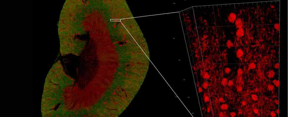
The device is not particularly large, but it can take exceptional pictures.
Image: ZEISS
limits of optics
But that’s where the research team at Zeiss came up against its limits: if the laser beam that generates the light sheets is focused even more sharply than before, then it becomes too short for the light sheet microscope. The laws of optics dictate this. “To overcome this problem, you need special beam shapes. These are the grid light sheets. We outsmart the classic optics and ensure that the light sheets are thin and at the same time very long.”
Another challenge: biologists grow cells on glass surfaces. You can illuminate them well from below, but not from the side, as is done with light sheet microscopy, explains physicist Ralf Wolleschensky from the Zeiss team: “Then we have to tilt the arrangement and look at an angle through the glass bottom. And This leads to very strong image errors. They are so strong that imaging is no longer possible. And that’s why we ended up having to develop a completely new type of optics for this purpose.”
Technology simplified
Technically, all of this was very demanding for the researchers at Zeiss, to whom Jörg Siebenmorgen is the third member of the team. However, the development proposed by the German Patent Office for the Future Prize now also has commercial potential. At the same time as the visual improvement, handling has been significantly simplified. So far, a lot of self-construction was necessary in the laboratories for the light sheet microscopy systems. It took a physicist to adjust and a computer scientist to evaluate.
Now there is, so to speak, an off-the-shelf device. “It is half a meter by half a meter in size, about 40 centimeters high and fits on any laboratory bench,” says Kalkbrenner. “Of course, there are still some peripherals: a laser module that provides the laser radiation. There is of course a computer system for control and calculation. But it has become a relatively compact system for a high-end fluorescence microscope.”
Helping research on malaria parasites…
Around 30 of these devices have now been sold worldwide – for a steep price, as Ralf Wolleschensky says. “We’re already talking about a well-equipped single-family home.” But the special microscope can look deeper into living cells than any other. And it helps researchers all over the world in a very concrete way: For example, the working group led by Ethan Goddard-Borger in Melbourne was able to follow the development of the malaria parasite in detail.
Kalkbrenner adds: “The team was able to observe some key points in the very complex life cycle of the parasite, which has various stages of development from humans to mosquitoes and back to humans, for the first time in living human blood cells. And as a result, they were also able to identify certain proteins that are responsible for these processes The prerequisites are. These are possible starting points for therapy.”
… and in cancer therapy
A second example comes from the University of Würzburg, which has one of three high-end fluorescence microscopes in Germany. Markus Sauer is using it to research an immunotherapy against cancer at the Franconian university. He was able to explain exactly how the contact between an immune cell and the tumor comes about.
This also offers potential for more targeted treatment. It will be interesting to see what the further development of light sheet microscopy will reveal about living cells.