Status: 02/10/2023 02:43 am
Wilhelm Conrad Roentgen died a hundred years ago. By chance he discovered radiation, which was later named after him – a milestone for medicine.
By Ralf Kölbel and Lilly Zerbst, SWR
As is so often the case in science, it was an accidental discovery: On November 8, 1895, the physicist Wilhelm Conrad Röntgen discovered invisible rays in Würzburg that were to revolutionize medical diagnostics. In 1901 he received the first Nobel Prize in Physics.
Exactly 100 years ago, on February 10, 1923, the discoverer of X-rays died. It was not recognized until much later that these can also be dangerous.
This is how X-rays are produced
X-rays are high-energy electromagnetic waves. They are created, for example, when very fast electrons are slowed down. The higher the amount of energy released, the shorter the wavelength of the radiation emitted.
In the spectrum of electromagnetic radiation, high-energy X-ray waves with a wavelength in the nanometer range lie between the lower-energy UV radiation and radioactive gamma radiation.
X-rays also occur naturally: in addition to visible light, the sun emits a broad spectrum of invisible electromagnetic radiation, including in the X-ray range. The earth’s atmosphere absorbs a large part of these rays. This means that the radiation exposure on the earth’s surface remains harmless.
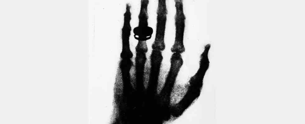
An accidental discovery
Roentgen had experimented with an almost airless glass cathode ray tube in which beams of electrons were bundled. He covered this tube with cardboard. However, the rays emitted by the electrons were able to penetrate the cardboard and revealed an object lying randomly on the table on the fluorescent screen.
At the end of 1895, Roentgen gave the Physical-Medical Society in Würzburg a first written communication “About a new type of radiation”. A little later he demonstrated his discovery publicly for the first time. He did not register a patent for this novelty. Because of this, the use of X-rays – as he himself called them – was able to spread quickly.
Wilhelm Conrad Roentgen received the award in physics at the first Nobel prizes being awarded.
Image: picture alliance / opale.photo
Look inside the body
In an X-ray machine as we know it today, electrons are released from a cathode by heating a filament and then accelerating the electrons. At the other end of the X-ray tube is an anode whose copper nuclei slow down the impacting electrons. The electrons give off just the right amount of energy to generate electromagnetic waves in the X-ray range.
The “X-ray”, later named after its discoverer, is used in medicine mainly to detect anomalies in the body. The tissues of the human or animal body of different densities absorb the X-rays to different extents. This allows you to look inside the body to detect broken bones or tumors, for example.
Further development of X-ray technology
The X-ray process has been further developed over the past few years. With modern X-ray computed tomography (CT), it is possible to capture three-dimensional images of the inside of the body. According to Professor Konstantin Nikolaou from the University of Tübingen, X-ray images of extremities can now be resolved with such a high resolution that the end result looks like a histological preparation of bone. The speed of technical progress in radiology cannot be compared to any other industry.
Artificial intelligence is now also being used: it helps to improve image resolution and thus better and better detect anomalies and diseases. The duration of the X-ray radiation, which is harmful to health, can also be reduced. In addition, new types of photon-counting computed tomographs are achieving ever higher resolution and thus greater diagnostic accuracy with shorter irradiation times and radiation doses.
Other radiation imaging techniques are also based on Roentgen’s discovery – including, for example, ultrasound examination, magnetic resonance imaging (MRT) and the radar method.
X-ray radiation health risk
For a long time, X-rays were used without thought. From 1920 onwards, many shoe shops around the world still had so-called “pedoscopes” as an attraction, with which one could check the fit of shoes using X-ray technology. The devices were particularly popular for children’s feet. Despite many indications of possible health hazards, they were only banned in Germany in 1973 by the Radiation Protection Act.
Although X-ray technology is useful, it should be used sparingly, especially in medicine. Because the high-energy X-ray waves can cause damage to the genetic material of the cells. This can also promote cancer. A high radiation dose also carries the risk of direct organ damage.
There is no scientifically recognized threshold value for X-ray exposure below which damage can be ruled out. In medical use, therefore, the following applies: An X-ray examination should only be ordered if the health benefit outweighs the risk.